Figure 2.
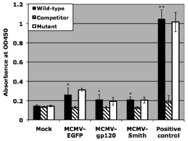
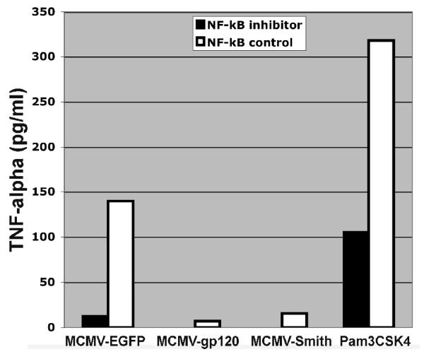
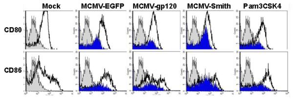
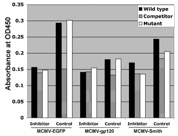
NF-κB activation is partly associated with DC activation by MCMV infection. A: NF-κB activation profile of MCMV-infected DC at 12 h after infection. Wild-type: NF-κB wild-type DNA. Competitor: NF-κB competitor DNA. Mutant: NF-κB mutant DNA. Positive control: HeLa Control Nuclear Extract. The experiment was repeated three times using Mo-DC prepared from three donors (Mean ± SD). * means p ≤ 0.05 compared to mock (t-test). ** means p < 0.001 compared to mock (t-test). B: TNF-α induction by MCMV-infected DC pre-treated with NF-κB inhibitor peptide or control peptide at 12 h after infection. Pam3CSK4 (1μg/ml), a known TLR2 ligand, serves as a positive control. The experiment was repeated twice and similar results were obtained. C: CD80 and CD86 expression by MCMV-infected DC pre-treated with NF-κB inhibitor peptide or control peptide at 48 h after infection. Gray filled histogram: PE labeled IgG1 isotype control antibody; Open histogram: Specific staining with either PE labeled CD80 or CD86 antibodies for control peptide treated cells. Blue filled histogram: Specific staining with either PE labeled CD80 or CD86 antibodies for NF-κB p65 inhibitor peptide treated cells. Pam3CSK4 serves as a positive control. Dead cells were excluded from the flow cytometric analysis by using propidium iodide staining. Representative results of two experiments are shown. D: NF-κB activation profile of MCMV-infected DC pre-treated with NF-κB inhibitor peptide or control peptide at 12 h after infection. Wild-type: NF-κB wild-type DNA. Competitor: NF-κB competitor DNA. Mutant: NF-κB mutant DNA. Representative results of two experiments are shown.


