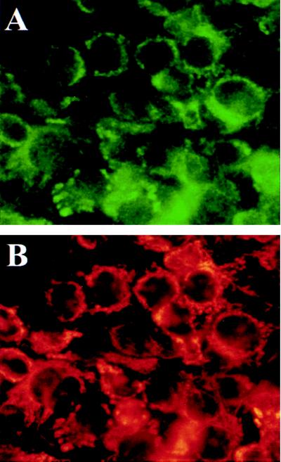
Figure 6.
WND protein is colocalized with mitochondria marker cytochrome c oxidase. Live HepG2 cells were incubated with selective mitochondria marker MitoTracker, and then cells were fixed and immunostained with aC-WND and secondary FITC-coupled antibody. Cells were photographed by using two filters to detect FITC fluorescence associated with the aC-WND staining (A) and the mitochondria-associated rodamine fluorescence of MitoTracker (B).

