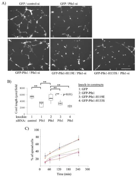
Figure 4. Cord morphogenesis of HmVEC requires functional interactions of Pfn1 with both actin and proline-rich ligands.
(A) Representative images of matrigel-induced cord formation by different groups of cells at 8 hrs after cell-seeding. (B) A box and whisker plot summarizing the cord morphogenesis data from a total of 2-3 independent experiments. (C) A line graph compares the relative spreading ability of different groups of cells at different time-points after seeding on matrigels (diamond: GFP/control-siRNA, cross: GFP/Pfn1-sRNA, triangle: GFP-Pfn1/Pfn1-siRNA, square: GFP-Pfn1-H119E/Pfn1 siRNA, and open circle: GFP-Pfn1-H133S/ Pfn1-siRNA). Data here are summarized from a total of two independent experiments with a duplicate set of samples for each experimental condition.

