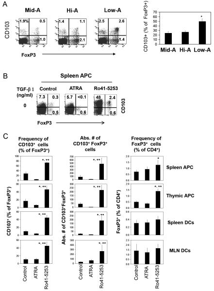
Figure 8. Increased production of CD103+FoxP3+ T cells in the thymus and by antigen presenting cells following RARα blockade.
(A) The frequencies of CD103+FoxP3+ T cells in the thymi of Mid-A, Hi-A or Low-A mice. (B) Induction of CD103+ FoxP3+ T cells by irradiated APC (prepared from the indicated organs of normal mice) in the presence of RO41-5253 or ATRA. Naïve CD4+CD25- T cells were co-cultured with irradiated APC or CD11c+ DCs for 5-6 days. One representative set of data out of at least 3 independent experiments are shown. The graphs in panel A (n=3) and C (n=4) show combined data with SEM. Significant differences from the control (*) or ATRA group (**).

