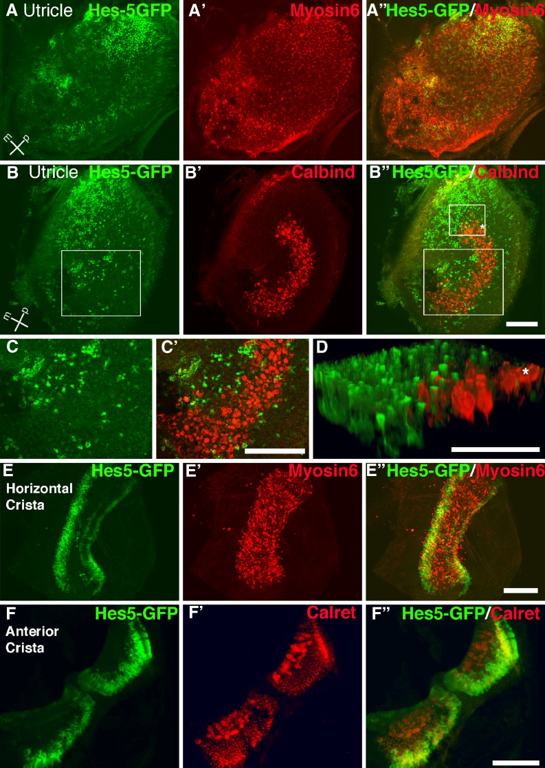
FIG. 5.
Hes5-GFP is expressed in a subset of adult vestibular supporting cells. Adult Hes5-GFP vestibular organs were immunolabeled for GFP and the hair cell marker Myosin6 (A–A” and E, E”), or the striolar (type I) hair cell calyx marker Calbindin (B–B”, C–C’, and D), or Calretinin (F–F”) which labels both hair cells and type I hair cell calyxes. Images are brightest point projections (A–A”, B–B”, C–C’, and E–E”) or 3D projections (D and F–F”) of confocal z-series micrographs. Hes5-GFP is expressed in a mosaic pattern in the adult utricle; there is an area of high expression in the medio-posterior region of the macula and scattered cells throughout the epithelium (A, B). Only a few Hes5-GFP labeled cells are found within the striolar region, labeled with anti-Calbindin (B–B”). The large boxed regions in B and B” are shown at higher magnification in C and C’, respectively. The smaller boxed region in B” is shown as an oblique 3D projection in D, rotated slightly counterclockwise; the radial profiles of Hes5-GFP labeled supporting cells can be seen at the margins of the striolar region, marked by the Calbindin-labeled striolar calyxes (asterisk). E–E” A horizontal crista shows Hes5-GFP labeling in supporting cells of the peripheral margins. F–F” The anterior crista has Hes5-GFP labeling surrounding the periphery of each half of the organ in a concentric pattern. Scale bar in B” = 100 μm and applies to A–A” and B–B”. Scale bars in C’, E”, and F” = 100 μm. Scale bar in D = 50 μm.

