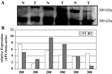
Fig. 5.
a Western blot analysis of APC protein expression in three normal (N) and tumor (T) tissue samples obtained from three patients with hepatocellular carcinoma. Two isoforms of the APC protein (300 and 200 kDa, respectively) were observed using antibodies directed against the APC protein. The sizes of corresponding marker fragments are shown on the right in kilodaltons. b Quantitative analysis of APC protein expression in hepatocellular cancer. The levels of APC expression in cancer and non-cancer tissues (the order of the patients is identical to the panel above) were standardized to the respective beta-actin expression levels. Overall no significant difference among the two isoforms was observed

