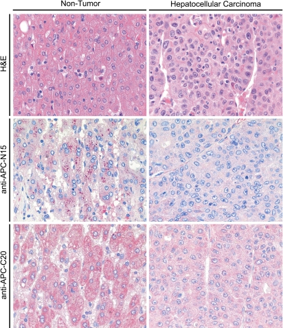
Fig. 6.
Immunohistochemical expression of APC in liver and hepatocellular carcinomas. The distribution and expression pattern of APC in non-tumorous liver and HCCs was investigated by immunohistochemistry. Non-tumorous epithelium, and tumor were stained with two different anti-APC antibodies (anti-APC-N15 and anti-APC-C20). APC was found in the cytoplasm. In the majority of the patients studied, the intensity of immunostaining and the number of immunoreactive cells was decreased in tumorous epithelium (right) when compared with the corresponding non-tumorous epithelium (left)

