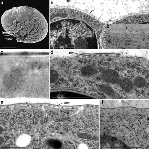
Fig. 2.
Extracellular matrix (ECM) before cuticle differentiation and the differentiation of embryonic cuticle I. a The stage 21 (S21) embryo is devoid of visible ECM. Scanning electron microscopy (SEM). b A thin amorphous ECM (arrow) covers the epidermal cells but is not labelled by gold-conjugated WGA (inset b’). c By contrast, chitin is detected in the basal half of the eggshell (black dots). d At S22, the apical plasma membrane forms microvillus-like structures (mv) at the tips of which the envelope (env) is produced. e Later, at S23, the envelope is continuous. f No chitin can be detected at this stage with gold-conjugated WGA. Transmission electron microscopy (TEM), cross sections. Bars 100 μm (a), 1 μm (b, d–f), 500 nm (c)

