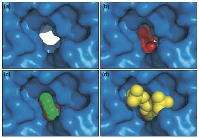
Figure 6.
Close-up view of the channel from the RNA binding side with (a) nothing bound; (b) modeled DMAPP, an Mg2+ ion, and three water molecules coordinated to the Mg2+ ion; (c) everything in (b) and the modeled base of A37; (d) everything in (c) and the modeled ribose and 5'- and 3'-phosphates of A37. DMATase is depicted in a surface representation and is in the same orientation as in Fig. 3(d). The molecules in the channel are in spheres, with DMAPP and water molecules in red, Mg2+ in magenta, base A37 in green, and the ribose and phosphates of A37 in yellow.

