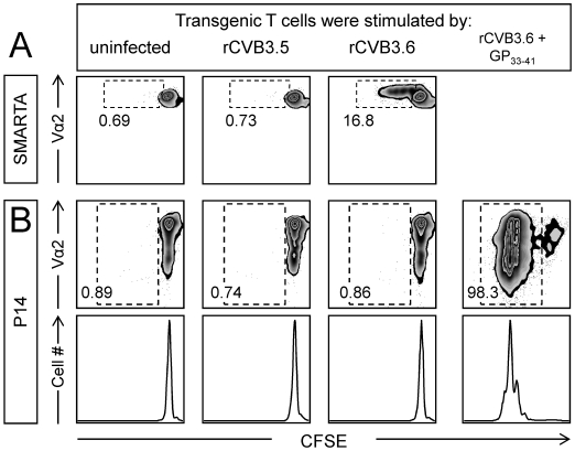
Figure 6. rCVB3.6 drives the in vitro proliferation of CD4+ T cells, but not of CD8+ T cells.
Stimulator cells: Splenocytes from wt C57BL/6 mice were left uninfected, or were infected with rCVB3.5 or rCVB3.6 (moi = 10). Some splenocytes (uninfected or rCVB3.6-infected) were pulsed with GP33–41 peptide. Indicator cells: P14 and SMARTA splenocytes were CFSE-labeled, then mixed to generate a 1∶1 ratio of both types of transgenic T cell. Indicator cells were added to wells containing stimulator cells and, 72 hours later, wells were harvested and analyzed by flow cytometry using FloJo. The panels shown are gated on CD8+/Thy1.1+ cells (P14) or CD4+/CD45.1+/Vα2+ cells (SMARTA). P14 cells were not gated on Vα2 because this molecule is down-regulated on activated/dividing P14 cells (as shown in the peptide-stimulated population in panel B); had we gated on Vα2hi cells, we would have failed to detect a large proportion of dividing cells. All analyses were done in triplicate, and the data shown are representative. The numbers are the proportion of (A) SMARTA cells or (B) P14 cells that have divided at least once, expressed as a percentage of total number of SMARTA or P14 transgenic T cells that were present in the plots.

