
Figure 1.
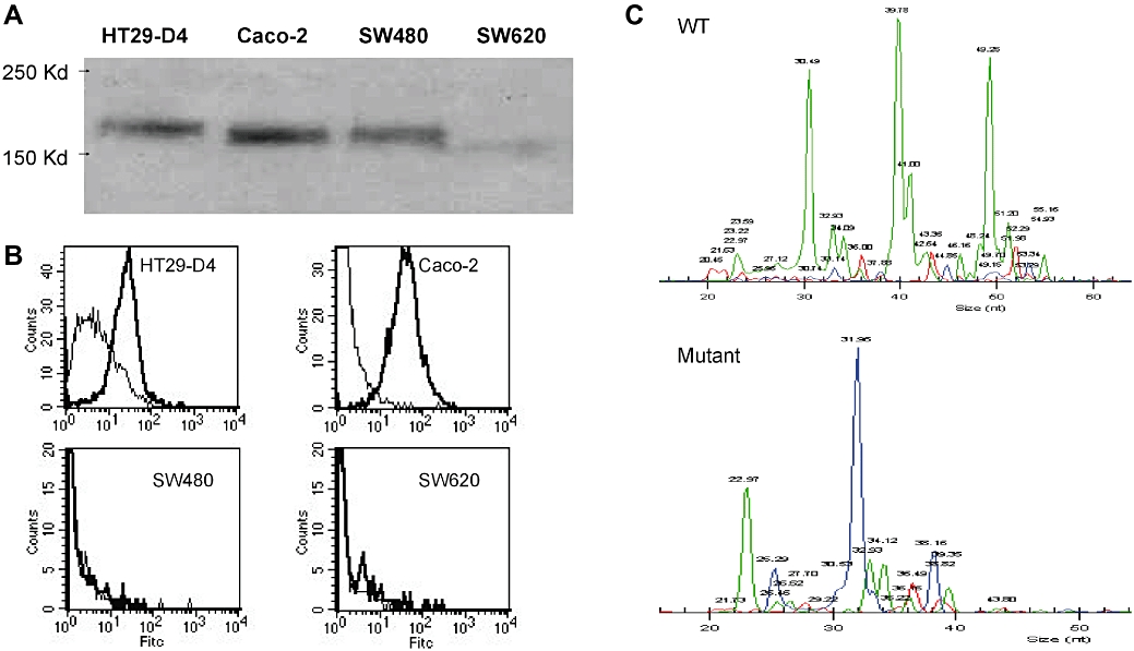
(A) Epidermal growth factor receptor protein expression was detected by immunoblotting cell lysates from four colon cancer cell lines HT29-D4, Caco-2, SW480 and SW620. (B) Epidermal growth factor receptor cell surface expression was measured by flow cytometry. Cells (5 × 10⁵) were incubated with cetuximab as primary antibody and counterstained with an Alexa Fluor 488 goat anti-human IgG. All staining were done on ice for 45 min followed by three washes. For each cell line, a control without primary antibody was performed. (C) Detection by SNaPShot of K-Ras mutations on cell lines. Each peak corresponds to a specific extended primer. Wild type (WT) for HT29-D4 and Caco-2 (upper panel); K-Ras mutation for SW480 and SW620 (lower panel).