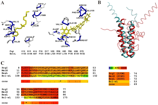
Figure 2. Active Site Similarity between PrgI and Bcl-xL.
(A) CPASS alignment of the S. typhimurium PrgI active-site complexed to DDAB with the active-site of human Bcl-2 protein (Bcl-xL) complexed with acyl-sulfonamide-based inhibitor. The residues aligned by CPASS are labeled and colored blue in the structures. The active site sequence alignment is also shown below the structures. The ligands are colored yellow. (B) Overlay of the human Bcl-2 protein (red) with S. typhimurium PrgI (turquoise) based on a DaliLite [29] alignment. (C) Multiple-sequence alignment of the three known T3SS structures of S. typhimurium PrgI, B. pseudomallei BsaL, and S. flexneri MxiH with the human Bcl-2 protein (Bcl-xL). The reliability of the each amino acid alignment is color-coded from blue (poor) to red (good) using the CORE index [35]. The consensus alignment received a score of 69, where a perfect alignment receives a score of 100.

