
Figure 1.
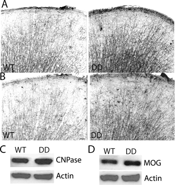
Enhanced myelination in Plp-Akt-DD mice. A, B, Coronal sections of cerebrum from 2-month-old WT and Plp-Akt-DD mice stained with antibodies against myelin proteins CNPase (A) and MOG (B). Note enhanced expression of both myelin proteins in Plp-Akt-DD brain relative to WT brain. A minimum of three animals were analyzed per genotype, and representative images are shown. Scale bar, 100 μm. C, D, Western blot data showing greater expression of CNPase (C) and MOG (D) in Plp-Akt-DD mice compared with WT mice at 2 months of age. CNPase expression relative to actin was 1.20 ± 0.08 (Plp-Akt-DD) compared with 0.97 ± 0.04 (WT); MOG expression relative to actin is 1.01 ± 0.11 (Plp-Akt-DD) compared with 0.71 ± 0.05 (WT). N = 3 and p < 0.05 in both cases.