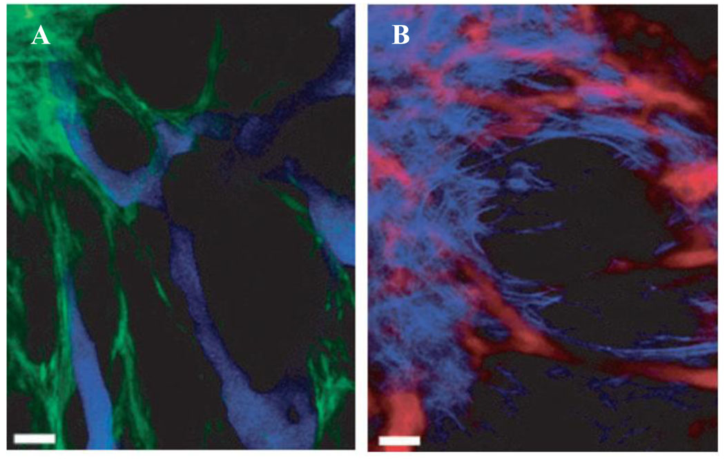
Figure 10.
Multi-photon microscopy represents a powerful tool for multiplexed in vivo imaging. By utilizing low-energy photons (minimally absorbed by tissues) for excitation of multicolor QD probes, this technique provides deeper tissue penetration and higher sensitivity of imaging. Application of this technique enabled the study of tumor morphology using QDs for labeling of tumor vasculature (blue QDs in (A) and red QDs in (B)), further enhanced by GFP labeling of perivascular cells (green in (A)) and detection of second harmonic generation signal from collagen to visualize extracellular matrix (light-blue in (B)) [58].

