
FIGURE 2.
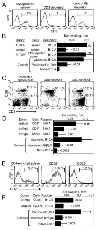
Regulatory cells from arrβgal mice are CD3+4+25+. (A, B) Analysis of CD3+ cells from arrβgal mice for Treg activity. (A) FACS analysis of CD3-depleted (center), and unfractionated spleen/LN cells (left). Percent of cells is indicated. (B) Depletion of CD3+ cells eliminates the ability to inhibit DTH upon transfer to B10.A mice. 30 × 106 cells were transferred. (C, D). Analysis of CD4+ cells from arrβgal spleen/LN for Treg activity. (C) FACS analysis of fractions selected for CD4+ cells (right), CD8+ cells (center), and unfractionated spleen/LN (left). (D) CD4+ cells transferred inhibition of DTH to B10.A mice. 7 × 10⁶ CD4+ or CD8+ T cells, or 30 × 106 unseparated cells were transferred to normal B10.A mice. (E, F). Transfer of CD25-depleted cells failed to inhibit the βgal ear swelling response. (E) FACS analysis of spleen/LN cells depleted of CD25+. Different clones of anti-CD25 were used for depletion (PC-61) or detection (7D4). 50 × 106 CD25- cells or 30 × 106 unseparated cells were transferred. (F) CD25- cells did not inhibit ear swelling. For all assays, the cells were transferred to B10.A mice on day 0, mice were immunized on day 1 by vaccination with VSC 56, and ear tested on day 26 - 28 with βgal. P values (t test) for comparisons with vaccinated B10.A control groups are given. Error bars indicate SD.