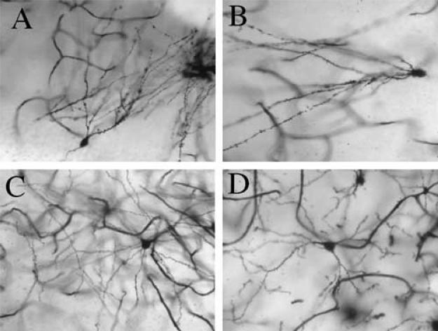
Fig. 1.
Displayed are representative photomicrographs of (A and B) dentate gyrus (DG) granular cells and of (C and D) nucleus accumbens (NAcc) pyramidal cells from animals treated neonatally with SAL (A and C) or MA (B and D). For the DG, spine density was decreased in MA animals (B) relative to SAL (A) animals; in the NAcc, dendritic branch length as well as spine density was decreased in MA animals (D) compared to SAL animals (C). It is important to note that these pictures do not represent the entire cell, as dendrites that course through tissue are not all on the same plane; however, the pictures depicted here are within the same plane of tissue. Pictures were taken with a Sony digital camera equipped with a Zeiss lens, and all pictures were taken at the drawing power for dendritic length (250×).

