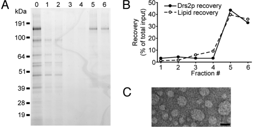
Fig. 4.
Flotation of TAP-Drs2p proteoliposomes in a glycerol gradient. (A) Fractions collected from the glycerol gradient were subject to SDS/PAGE, and the gel was stained with SimplyBlue. Lane 0, the protein sample before reconstitution; lanes 1–6, fractions (#1–6) collected from the bottom to the top of the gradient. (B) Recovery of Drs2p and phospholipid in each fraction relative to the starting material. (C) Electron micrograph of negative-stained proteoliposome sample from fraction #5. (Scale bar, 50 nm.)

