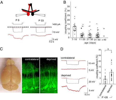
Fig. 1.
Sensory deprivation prevents the developmental decrease in coupling coefficients and alters olfactory bulb development. (A) Diagram of whole-cell somatic recordings from pairs of mitral cells that project to the same glomerulus. Hyperpolarizing current injections (range −250 to −400 pA, 0.7 s) into cell 1 (black) elicited a voltage deflection in the cell 2 (red). Each trace represents an average of 7–10 sweeps. Mitral cells at P8 showed higher coupling than at P33. (B) The coupling coefficient (ratio of the hyperpolarization in the test/stimulated cell) decreased after P10. (C) The right olfactory bulb was smaller in this P32 mouse after right naris occlusion at P1. The laminar organization of the olfactory bulb and the projection of apical dendrites to glomeruli were not altered in sensory-deprived animals. The images show slices from contralateral and sensory-deprived bulbs (P > 28), in which Thy1-YFP is expressed in a proportion of mitral cells. (Scale bar, 50 μm.) (D) In young adult mice (P > 28), the coupling coefficient was greater on the side that was sensory deprived. In the example contralateral control bulb shown, a hyperpolarizing current injection (−400 pA, 0.7 s) into cell 1 (black) elicited only a small voltage deflection in cell 2 (red), compared with the larger voltage deflection elicited in the deprived bulb (P < 0.001).

