Fig. 2.
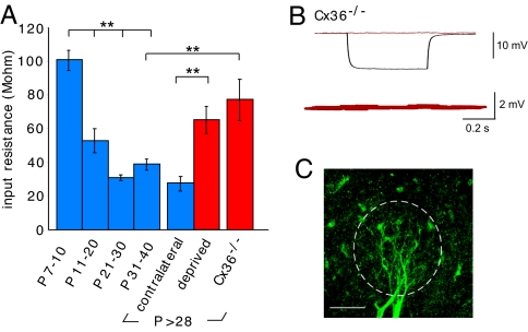
Sensory deprivation and genetic deletion of Cx36 prevent developmental changes in membrane conductance. (A) Input resistance decreased in control mitral cells for all time intervals after P7–10 including the contralateral control group (P < 0.001). In contrast, input resistance for the sensory-deprived and Cx36−/− animals was much higher than the contralateral bulbs or age-matched controls, respectively (P < 0.001, P < 0.002). (B and C) As expected, in Cx36−/− animals a current pulse caused a hyperpolarization in one cell (black trace), but no electrical coupling in the follower cell (red trace). The confocal image shows 2 biocytin-filled mitral cells from a Cx36−/− mouse (P34). The margin of the glomerulus is outlined by the dotted line. (Scale bar, 50 μm.)

