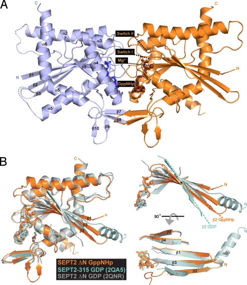
Fig. 1.
Structure of SEPT2·GppNHp. (A) Ribbon model of the overall structure of the SEPT2 dimerized across the nucleotide-binding site. New elements observed are labeled and the GppNHp and magnesium are colored in brown. (B) Superimposition of the SEPT2·GppNHp structure (gold) with the previous structures of SEPT2·GDP [PDB ID 2QA5 (cyan) and 2QNR (gray)].

