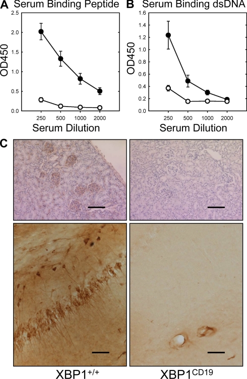
Figure 1.
Female XBP1+/+ and XBP1CD19 mice were immunized i.p. with 100 µg MAP-DWEYS in CFA followed by booster immunizations with 100 µg MAP-DWEYS in IFA on days 14 and 28. (A and B) On day 35, serum was tested for anti-DWEYS (peptide; A) and anti-dsDNA (B) antibodies by ELISA of four serum dilutions. In both panels, each circle represents the mean ± SEM value for XBP1+/+ (closed circles) or XBP1CD19 (open circles) mice. (C) XBP1+/+ and XBP1CD19 mice from A and B were treated i.p. with 3 mg/kg LPS on days 52 and +54. Shown are representative images of IgG-specific IHC of kidney (top) and hippocampus (bottom) of mice 4–7 d after LPS treatment. XBP1+/+ mice demonstrated anti-IgG binding to neurons in the CA1 region of the hippocampus. There was no anti-IgG binding in any of the XBP1CD19 animals. Bars: (kidney) 200 µm; (hippocampus) 100 µm. All panels represent results from a single experiment with five mice per group. A second independent experiment reproduced these findings (not depicted).

