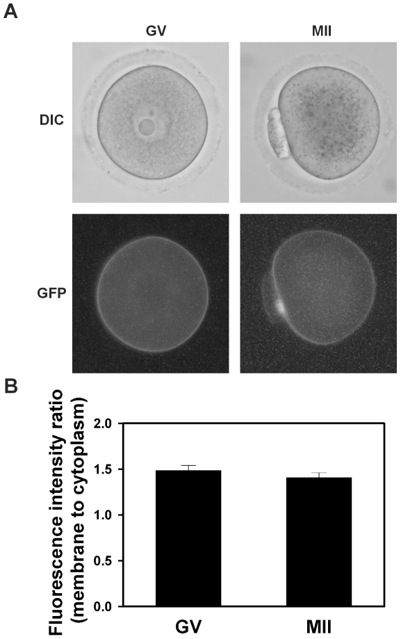
Figure 7. Localization of hD1-GFP in mouse oocytes during meiotic maturation.
(A) Representative example of hD1-GFP-expressing GV oocyte and MII egg as indicated at top showing fluorescence (lower panel) and corresponding phase-contrast image (upper panel). (B) Mean (±s.e.m.) plasma membrane-to-cytoplasm fluorescence ratios of hD1-GFP-expressing GV oocytes and MII eggs (N = 4, with 17 images analyzed in each group; not significantly different by student's two-tailed t test).

