Fig. 1.
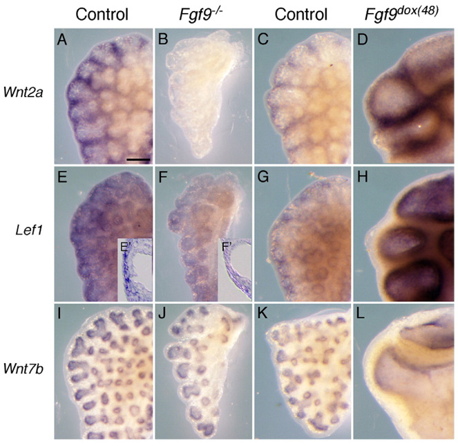
FGF9 regulates WNT signaling in lung mesenchyme. (A–H) Whole-mount in situ hybridization showing Wnt2a (A–D) and Lef1 (E–H) expression in E13.5 lung. All tissue pairs were hybridized and developed together in the same tube. Comparison of control (A) and Fgf9−/− (B) lungs shows an absence of Wnt2a expression in lung mesenchyme in Fgf9−/− tissue. In contrast, induced overexpression of FGF9 in lung epithelium from E11.5 to E13.5 (Fgf9dox(48)) (D) results in increased Wnt2a expression compared with the control lungs (C). Comparison of control (E) and Fgf9−/− (F) shows decreased expression of Lef1 in Fgf9−/− lung mesenchyme. Comparison of control (G) and Fgf9dox(48) (H) lungs shows increased expression in FGF9 overexpressing mesenchyme. Cryo-sections (inset) of Lef1 whole-mount in situ hybridization stained control (E′) and Fgf9−/− lungs (F′) reveal decreased Lef1 expression localized in the sub-mesothelial mesenchyme. Color development in panels C, D, G, H were for shorter periods of time compared to panels A, B, E, F. (I–L) Wnt7b expression levels in lung epithelium was not affected in either FGF9 loss of function (Fgf9−/−, J) or gain of function (Fgf9dox(48), L) lungs compared with the controls (I, K). Scale bar in panel A, 200 µm.

