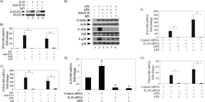
FIGURE 4.
IL10 antagonism does not impair the anti-inflammatory actions of adiponectin in human macrophages stimulated with LPS. A, cells were incubated with no addition (lanes 1 and 2), 10 μg/ml anti-IL10 antibody (lane 3), or control IgG (lane 4) for 18 h and subsequently stimulated with (lanes 2–4) or without (lane 1) 10 ng/ml IL10 for 30 min. Whole-cell lysates were fractionated by SDS-PAGE and immunoblotted with antibodies to phospho-STAT3. Total STAT3 served as loading control. B and C, cells were treated with or without 10 μg/ml adiponectin for 18 h in the presence of anti-IL10 antibody or control IgG as indicated and subsequently stimulated with 5 ng/ml LPS for 6 h. RT-qPCR measured the levels of IP10 (B) and TNFα (C) mRNAs, using 18S as internal control for adjustment between samples. Data are expressed in means ± S.E. (n = 3) relative to the values of samples from cells without adiponectin or LPS treatment. *, p < 0.05. D, cells were incubated with (lanes 3–6) or without (lanes 1 and 2) 10 μg/ml adiponectin for 18 h in the presence of anti-IL10 antibody or control IgG as indicated and subsequently stimulated with 5 ng/ml LPS for 40 min. Whole-cell lysates were fractionated by SDS-PAGE and immunoblotted with antibodies to phospho-IκB (top), phospho-JNK (center), or phospho-p38 (bottom). Actin, total JNK, and total p38 served as loading controls, respectively. E, cells were transfected with IL10-specific or control siRNA as indicated. At 48 h after transfection, cells were incubated with or without 10 μg/ml adiponectin for 6 h. RT-qPCR measured IL10 mRNA levels using 18S as internal control for adjustment between samples. Data are expressed in means ± S.E. (n = 3) relative to the values of samples from cells transfected with control siRNA and left unstimulated. *, p < 0.05 versus control. F and G, cells were transfected with IL10-specific or control siRNA as indicated. At 48 h after transfection, cells were incubated with or without 10 μg/ml adiponectin for 18 h and subsequently stimulated with 5 ng/ml LPS for 6 h. RT-qPCR measured IP10 (F) and TNFα (G) mRNA levels using 18S as internal control for adjustment between samples. Data are expressed in means ± S.E. (n = 3) relative to the values of samples from cells transfected with control siRNA and left unstimulated.*, p < 0.05.

