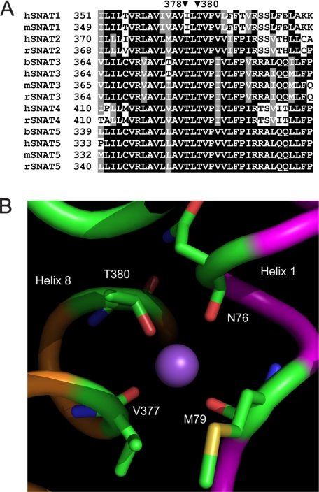
FIGURE 8.
Sequence alignment of members of the SNAT (SLC38) family and putative SNAT3 Na+-binding site. A, reference sequences for SNAT1–5 were downloaded and aligned using ClustalW. The alignment around rat SNAT3 residue Thr-380 is shown. The corresponding positions in other family members are indicated. Human, mouse, rat, and bovine sequences are indicated by prefix h, m, r, and b, respectively. B, close view of the proposed Na+-binding site of SNAT3. Helix 1 is shown in purple, and helix 8 is in orange. Residues potentially involved in Na+ binding are indicated.

