FIGURE 3.
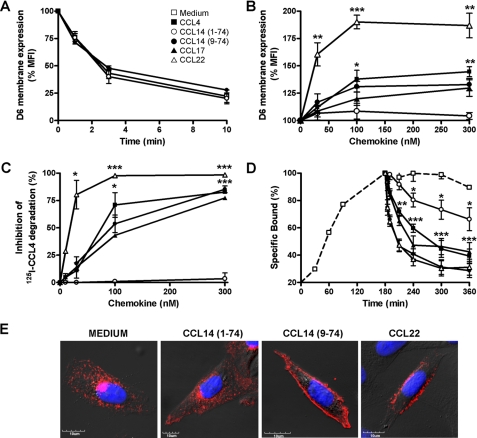
CCL14(1–74) is not a D6 antagonist. A, D6 internalization. CHO-K1/hD6 transfectants were incubated at 4 °C for 1 h with anti-D6 antibody. Cells were then incubated at 37 °C for different times in the presence of 116 nm of CCL4 (■), CCL14(1–74) (○), CCL14(9–74) (●), or only medium (□) and then stained with secondary antibody for 1 h at 4 °C. B, D6 membrane up-regulation. CHO-K1/hD6 transfectants were incubated at 37 °C for 1 h with increasing concentrations of CCL4 (■), CCL14(1–74) (○), CCL14(9–74) (●), CCL17 (▲), and CCL22 ( ) and then labeled with αD6-PE antibody. Results are expressed as the percentage of MFI basal D6 expression. C, inhibition of D6-mediated CCL4 scavenging. CHO-K1/hD6 transfectants were incubated for 3 h with 0.1 nm 125I-CCL4 and increasing concentrations of unlabeled CCL4 (■), CCL14(1–74) (○), CCL14(9–74) (●), CCL17 (▲), and CCL22 (
 ). Results are expressed as 100 minus the percentage of degraded radiolabeled chemokine present in the supernatant. D, 125I-CCL4 dissociation curves. CHO-K1/hD6 transfectants were incubated for 3 h at 4 °C with 50 pm 125I-CCL4, washed to remove unbound radioligand, and incubated for increasing times with 100 nm CCL4 (■), CCL14(1–74) (○), CCL14(9–74) (●), CCL17 (▲), and CCL22 (○) or only medium (□). Results are expressed as 100 minus the percentage of radioligand remaining cell-associated. Data shown in A–D are the mean ± S.E. of at least three independent experiments performed. *, p < 0.05; **, p < 0.005; ***, p < 0.0005, chemokine- versus medium-treated CHO-K1/hD6 transfectants. E, confocal images of immunofluorescence-stained CHO-K1/D6 cells. Shown is one representative experiment of at least three performed of D6 staining after stimulation with the indicated chemokines (100 nm, 30 min). Each panel shows the double staining of D6 (indicated in red) with 4′,6-diamidino-2-phenylindole (indicated in blue).

