
Figure 2. Syt utilizes C2B to bend membranes in a response to Ca2+.
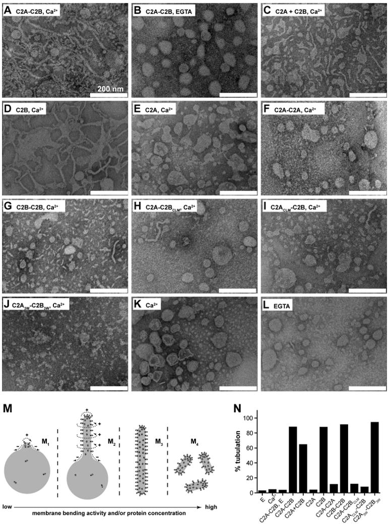
(A-L) Electron micrographs of Folch liposomes incubated with the indicated syt fragments (10 μM) for 10 hr. Scale bars correspond to 200 nm. The diameters of the lipid tubules were (mean ± SD, n = 30): 16 ± 4 nm for C2A-C2B; 11 ± 2 nm for C2B and 10 ± 3 nm for C2A3W-C2B3W. (M) Model showing that different degrees of membrane-bending may result in different morphologies of membranes. Membranes are shown in gray, syt fragments are shown as small ovals. Positive and negative curvatures are indicated as ‘+’ and ‘−’, respectively. Penetration of syt into the membrane drives deformation of the bilayer. The resulting morphologies can be classified into four categories, as follows: (M1), formation of a ‘nipple’, which has positive curvature at the tip, but negative curvature at the base, as indicated by dashed arrows. Protein prefers positively curved membranes and tends to cluster at the nipple tip. This in turn helps to maintain the shape of the nipple. (M2), more extensive membrane bending causes elongation of the nipple, forming a lipid tubule protruding from the parental liposome. New areas with a high degree of positive curvature emerge on the radial plane resulting in increased membrane area that is favorable for protein binding. (M3), even more extensive membrane bending would transform a whole liposome into a long tubule, removing any negative curvature. (M4), in extreme cases, the long lipid tubules are broken into short tubules or even tiny vesicles, further increasing the area with positive curvature. (N) Quantification of the fraction of tubulated liposomes obtained in the presence of the indicated syt construct.