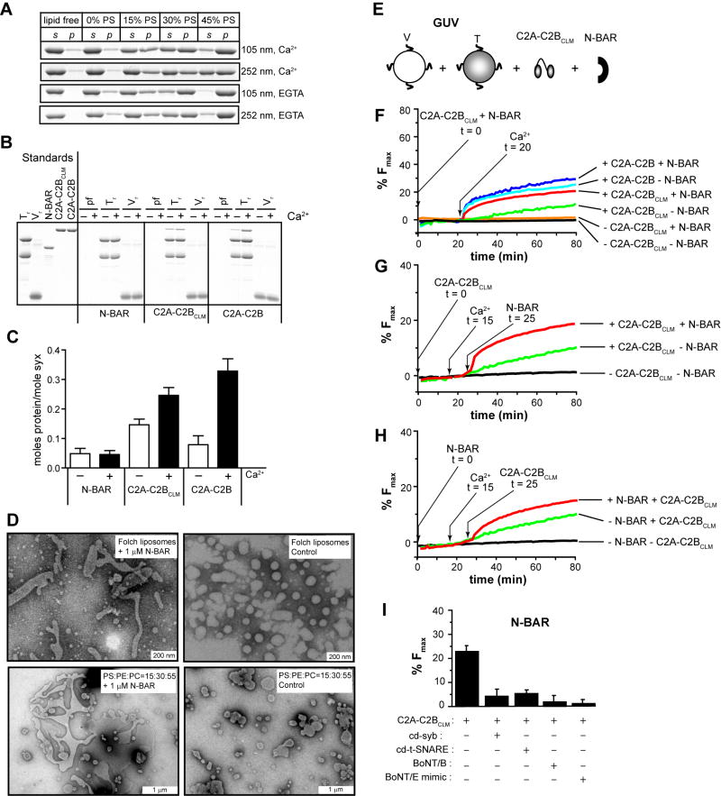
Figure 5. N-BAR domain induced membrane-bending rescues C2A-C2BCLM-regulated membrane fusion.
(A) Representative cosedimentation gels showing that N-BAR binds PS harboring liposomes in a Ca2+-independent manner. With increasing levels of PS, N-BAR shifted from the supernatant (s) to the pellet (p) fraction. At the same %PS, N-BAR binds more avidly to V105nm than to V252nm. (B) N-BAR does not exhibit significant t- or v-SNARE binding activity, while C2A-C2BClm and C2A-C2B bind t-SNAREs in a Ca2+-dependent manner, as assessed using coflotation assays. Protein free, t-SNARE- and v-SNARE-bearing vesicles are indicated as pf, Tr and Vr, respectively. (C) Quantification of coflotation assays. (D) Electron micrographs showing that 1 μM N-BAR tubulated liposomes composed of 100% Folch lipids or 15% PS/30% PE/55% PC, after 15 min incubation; the diameters of the tubules ranged from 30 nm to 90 nm. (E) In vitro membrane fusion assay setup. v-SNARE and t-SNARE GUVs were mixed with 10 μM C2A-C2BClm and 1 μM N-BAR. (F) Addition of N-BAR greatly enhanced both the rate and extent of Ca2+-triggered C2A-C2BCLM-stimulated GUV-GUV fusion, but had modest effects on WT C2A-C2B-stimulated fusion; in the absence of N-BAR, C2A-C2BCLM-stimulated GUV-GUV fusion was negligible (green trace). 1 mM Ca2+ was added at t = 20 min. (G) Sequential addition of 10 μM C2A-C2BCLM (t = 0), 1 mM CaCl2 (t = 15 min), and 1 μM N-BAR (t = 25 min) to GUV t-SNARE and v-SNARE vesicles, as indicated by arrows. (H) Sequential addition experiment as carried out in C, with N-BAR added at t = 0 and C2A-C2BCLM added at t = 25 min, as indicated by arrows. (I) N-BAR-rescued fusion was inhibited by the same agents used in Figure 4F.

