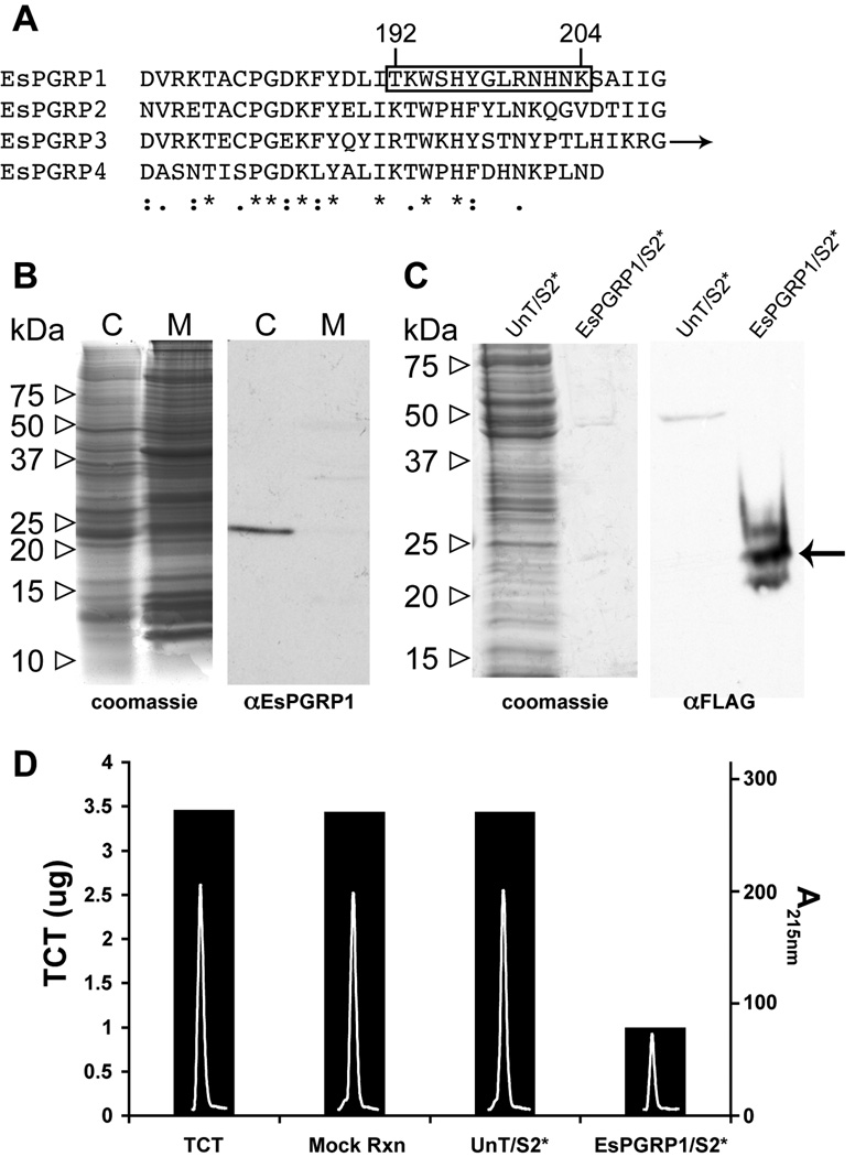
Figure 2. Biochemical characteristics of the EsPGRP1 protein.
A) An alignment of the EsPGRP C-termini (arrow indicates additional C-terminal sequence for EsPGRP3). Boxed area highlights antigen sequence used for anti-EsPGRP1 antibody generation. *, identical residues; :, conserved substitutions; ., semi-conserved substitutions. B) A representative immunoblot of E. scolopes tissue. The anti-EsPGRP1 antibody reacts with a major band in the aqueous soluble fraction at ~24 kDa. C, cytoplasmic (aqueous) fraction; M, membrane (SDS soluble) fraction. C) Anti-FLAG immunoblot and companion gel of 24 µg crude protein extract from untransfected Drosophila S2* cells and 256 ng of an EsPGRP1-enriched fraction of S2* cells transfected with an EsPGRP1-FLAG expression construct. The arrow indicates an anti-FLAG reactive band at the expected molecular weight for EsPGRP1. D) An EsPGRP1 enriched EsPGRP1/S2* fraction degraded TCT in vitro, but a protein extract of untransfected S2* cells had no effect on TCT levels. Bars indicate mass of TCT (left axis) in each reaction, white peaks inside bars are the TCT peaks following treatment as the absorbance at 215nm on a reverse-phase HPLC chromatograph (right axis). UnT/S2*, untransfected S2* cell extract +; EsPGRP1/S2*, EsPGRP1-enriched fraction of S2* cells transfected with pJT28; mock rxn, TCT incubated in reaction buffer for 3 h; TCT, 7 µL 1mM TCT loaded directly onto HPLC column.

