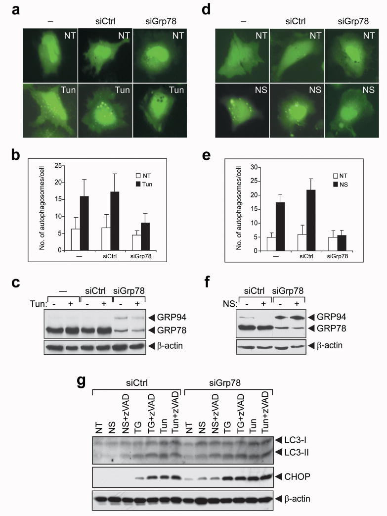
Figure 5.
GRP78 knockdown blocks autophagosome formation induced by ER stress or NS. (a) HeLa cells were transiently transfected with GFP-LC3 alone (−) or in combination with 40 nM of siCtrl or siGrp78. The cells were then either non-treated (NT) or treated with Tun (3 μM) for 4 h. Representative images of GFP-LC3 punctate dot formation in live cells captured by fluorescence microcopy are shown. (b) Quantitation of autophagosome formation in cells treated as in (a). The results from 3 independent experiments were summarized and expressed as the mean with the indicated SD. (c) Western blot analysis of GRP78, GRP94 and β-actin (as loading control) protein levels in cells treated as in (a). (d–f) Same as (a–c) except the cells were either non-treated (NT) or subjected to NS. (g) HeLa cells were transfected with 40 nM of either siCtrl or siGrp78 and subjected to NS treatment for 2 h, TG or Tun treatment for 4 h in the absence or presence of zVAD-fmk (40 μM) as indicated on top. The levels of LC3-I and II, CHOP and β-actin were detected by Western blot. The experiments were repeated 2 times.

