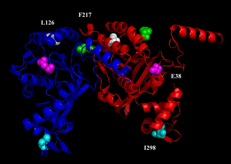
Figure 1.

This figure shows a depiction of two adjacent RecA molecules (red and blue) as they may appear in a helical filament based on the crystal structure without DNA (Story and Steitz, 1992; Story et al., 1992). The green residues represent the positions of F217Y, the magenta residues show the positions of E38K, the white residues are L126V and the cyan residues are I298V.
