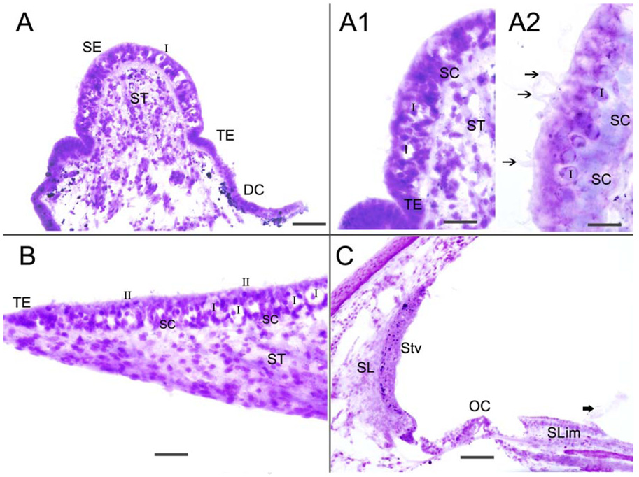
Fig 1.
Hematoxylin and eosin stained frozen section of the crista ampullaris (Fig 1A-A2), macula utricle (Fig 1B) and cochlea (Fig 1C). Fig 1A shows the crista sensory epithelia (SE), the transitional epithelia (TE), the dark cell area (DC) and the stroma tissue (ST). Fig 1A1 and 1A2 are higher magnification views of the cristae. The sensory epithelia is well-preserved and type I (I) and type II (II) hair cells, as well as supporting cells (SC), and cells in the stroma (ST) are easily identified. Hair cell stereocilia can be seen (arrowheads). Fig 1B shows the macula utricle which exhibits good preservation of the sensory epithelia hair cells and supporting cells, with mild swelling of calyceal terminals that surround type I hair cells (I). Figure 1C is a cross-section of the cochlea; there is good preservation of the organ of Corti (OC), spiral ligament (SL) and stria vascularis (Stv) as well as the spiral limbus (Slim). In this specimen, the tectorial membrane and Reissner’s membrane had become detached during microdissection (thick arrowhead). Magnification bar is 100 µm for (A) and (C), 50 µm for (A1), 20 µm for (A1), 60 µm for (B).

