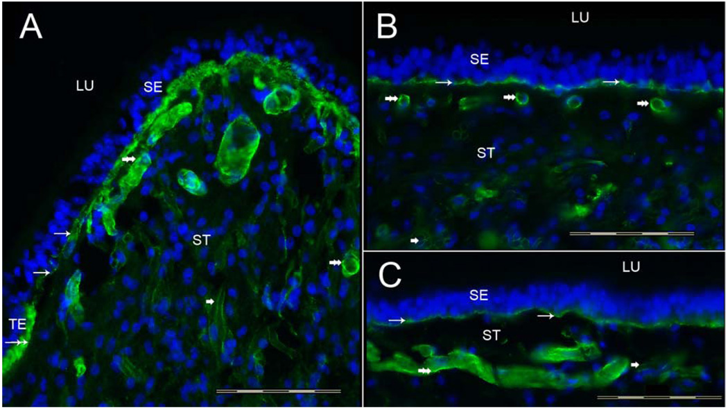
Fig 2.
Collagen IVα2 immunoreactivity (-IR) in the human vestibular endorgans. Fig 2A shows a cross-section of the crista ampullaris, close to the central region. Collagen IVα2-IR (green color) was seen within the BM underneath the sensory epithelia (SE) (thin arrows). The BM underneath the transitional epithelia (TE) also displayed collagen IVα2-IR (double arrow). The perivascular BMs within the underlying stroma (ST) were collagen IVα2-IR (double arrowheads). The perineural BMs beneath the epithelia and within the underlying ST also exhibited collagen IVα2-IR (single arrowhead). The macula utricle (Fig 2B) and macula saccule (Fig 2C) showed a similar collagen IVα2-IR pattern. BMs that surround stromal myelinated nerve fibers (arrowhead) and blood vessels (double arrowhead) of the maculae utricle and saccule were also collagen IVα2-IR. DAPI (blue color) identifies cell nuclei. LU: Lumen. Magnification bar is 200 µm for all figures.

