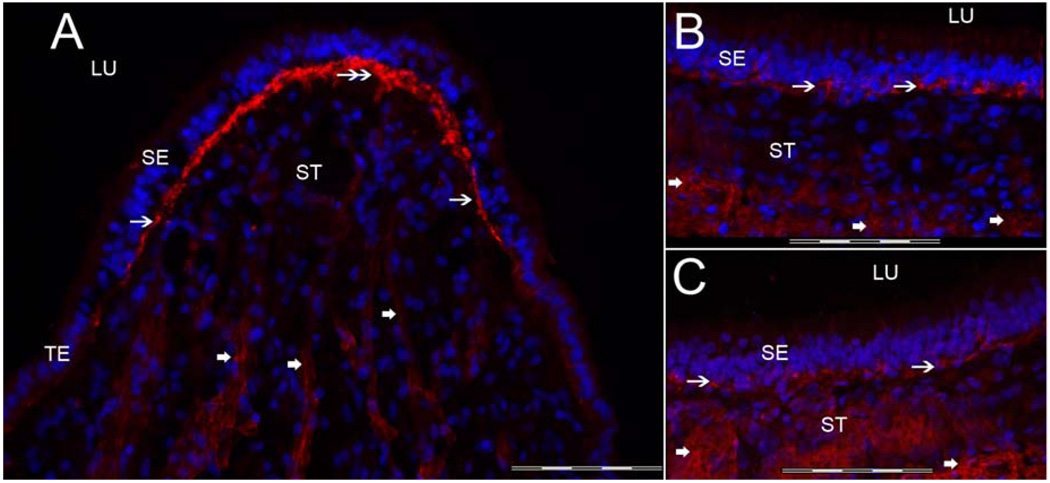
Fig 6.
α-Dystroglycan-IR. Fig 6A is a cross-section of the crista ampullaris, close to the central zone. α-dystroglycan-IR (red color) was seen within the BMs underneath the sensory epithelia (SE) (double arrows). In comparison to the BM proteins, collagen IV and laminin-β2, the α-dystroglycan-IR was relatively less intense at the lateral and basal zone of the crista ampullaris (single arrow), and significantly less prominent within the BMs beneath the TE and within the perivascular BMs. The macula utricle (Fig 6B) and macula saccule (Fig 6C) exhibited a similar α-dystroglycan-IR pattern as the crista ampullaris. α-dystroglycan-IR was noted in BMs underneath the maculae sensory epithelia in close apposition with the basal portion of the supporting cells (arrows). BMs that surround stromal myelinated nerve fibers of the macula utricle and saccule also exhibited α-dystroglycan-IR (thick arrowheads). DAPI (blue color) identifies cell nuclei. Magnification bar is 200 µm for all figures.

