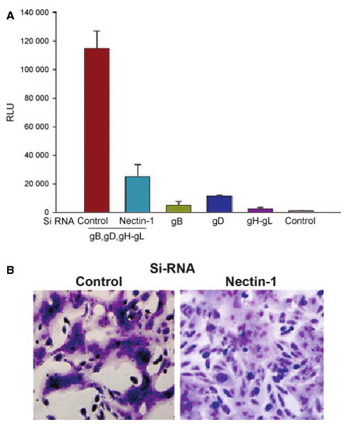
Fig. 6.

Role of nectin-1 in HSV-1-induced fusion of RPE cells. (A) Membrane fusion of RPE cells requires nectin-1 and the presence gB, gD, gH and gL. The ‘target’ RPE cells were transfected with either a control or nectin-1-specific siRNA. The ‘effector CHO-K1 cells’ were transfected with expression plasmids for the HSV-1 glycoproteins indicated, and mixed with ‘target RPE cells’. Membrane fusion as a means of viral spread was detected by monitoring luciferase activity. Relative luciferase units (RLUs), determined using a Sirius luminometer (Berthold detection systems), are shown. Error bars represent standard deviations. *P < 0.05, one-way ANOVA. (B) Downregulation of nectin-1 inhibits HSV-1-induced cell-to-cell fusion. The ‘effector CHO-K1 cells’ were mixed with either control (B, left panel) or nectin-1-specific siRNA-transfected (B, right panel) ‘target RPE cells’. At 18 h postmixing, the cells were fixed and stained with Giemsa to demonstrate syncytia formation.
