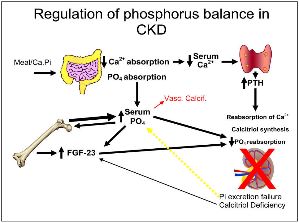
Figure 2.
Regulation of phosphorus balance in CKD. Meal associated Ca and PO4 are absorbed. Decreased Ca absorption and hypocalcemia stimulate parathyroid hormone secretion. Absorbed PO4 is deposited in the skeleton through bone formation or excreted by the kidney. Skeletal osteocytes read bone formation, and when the available PO4 exceeds skeletal deposition (bone formation), they secrete FGF23 to have the kidney excrete the excess PO4. In CKD, renal excretion of PO4 fails to maintain balance despite PTH and FGF23 influence and positive PO4 balance results (yellow arrow) and the serum PO4 begins to rise. This is a direct stimulus to heterotopic mineralization (red arrow and vascular calcification as a form of heterotopic mineralization).

