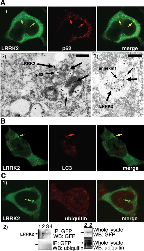
Figure 4.
A proportion of LRRK2 puncta colocalize with the proteins p62 and LC3, but not with ubiquitin. (A) LRRK2 puncta colocalize with p62 endogenous puncta. BAC-YPet-LRRK2-WT was transfected in HEK-293 cells and double-labelling performed by IF (A1) or IEM (A2 and 3) using anti-GFP (thick arrows in A2 and 3) and either anti-p62 [A1 and 2 (thin arrows in A2)] or anti-annexin1 (A3) antibodies (thin arrows). Scale bar in image A2 represents 200 nm although in A3 represents 500 nm. (B) LRRK2 puncta colocalize with LC3 protein. BAC-YPet-LRRK2-WT was transfected in HEK-293 cells stably expressing HA-mCherry-LC3 and analysed by IF using anti-HA and anti-GFP antibodies. (C) BAC-YPet-LRRK2-WT, BAC-YPet-LRRK2-G2019S or BAC-YPet-LRRK2-R1441C were expressed in transfected cells and double labelling performed using YPet-LRRK2 fluorescence and anti-ubiquitin antibodies (BAC-YPet-LRRK2-WT in C1). Alternatively protein lysates were prepared and subjected to immunoblotting either directly or after immunoprecipitation using anti-GFP antibodies (C2). Legend of samples: 1 = Untransfected, 2 = BAC-YPet-LRRK2-WT, 3 = BAC-YPet-LRRK2-G2019S and 4 = BAC-YPet-LRRK2-R1441C. A1, B and C1 are confocal images.

