Figure 1.
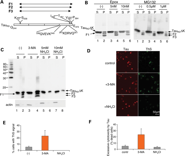
Effects of NH4Cl and 3-MA on the cleavage, aggregation and cytotoxicity of TauRDΔK in N2a cells. (A) Diagram of TauRD showing cleavage sites and CMA recognition motifs. TauRD is first cleaved after K257 to generate F1, then after V363 to generate F2, and further after I360 to produce F3. The two CMA recognition motifs are 336QVEVK340 and 347KDRVQ351. (B and C) Blot analysis of effects of proteasomal inhibitors (epoxomicin or MG132) (B) or autophagy–lysosomal inhibitors (3-MA or NH4Cl) (C) on cleavage and oligomerization of TauRDΔK. N2a cells overexpressing TauRDΔK were treated with these inhibitors for 5 days. Lanes labeled P (=pellet) denote sarkosyl-insoluble Tau species, S (=supernatant) indicates soluble proteins. Note that NH4Cl but not 3-MA inhibits the cleavage and aggregation of TauRDΔK. (D) ThS staining showing the influence of 3-MA and NH4Cl on Tau aggregation. (E) Quantification of ThS positive cells, which increase ∼5-fold with 3-MA but not with NH4Cl. (F) Effects of NH4Cl and 3-MA on the cytotoxicity of TauRDΔK in N2a cells. The excessive cytotoxicity by Tau was obtained by subtracting cytotoxicity without Tau from cytotoxicity with Tau expression. Note that 3-MA but not NH4Cl aggravates TauRDΔK cytotoxicity.

