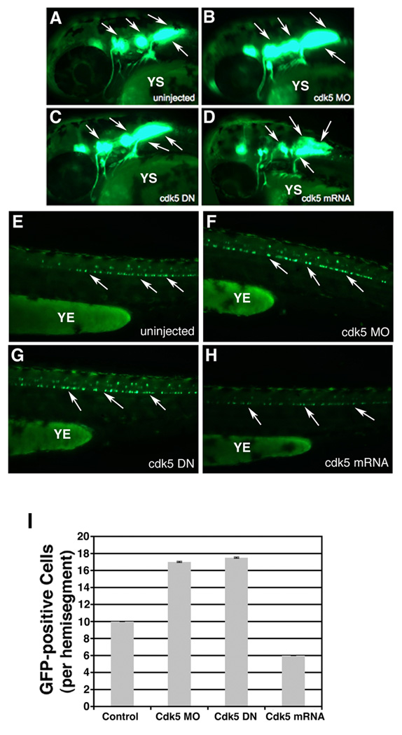
Figure 3. Zebrafish embryos at 48 hpf continue to show over-production of motor neurons upon cdk5 knockdown.
Images of live 48 hpf islet 1:GFP transgenic fish. Lateral views of the anterior regions of the embryos show motor neurons in (A) uninjected control, (B) cdk5 morpholino (MO)-injected, (C) kinase-dead cdk5 (human) mRNA-injected and (D) zebrafish cdk5 mRNA (50)-injected embryos. Arrows indicate the motor neuron populations in the brain. YS indicates yolk sac. Lateral views of the posterior region of the embryos show GFP-positive spinal motor neurons in, (E) uninjected control, (F) cdk5 morpholino (MO)-injected, (G) kinase-dead cdk5 (human) mRNA-injected (cdk5 DN), and (H) zebrafish cdk5 mRNA (50 pg injected/ embryo). YE indicates yolk extension. Arrows indicate some of the spinal motor neurons. The GFP-positive neurons or cells per each hemisegment are counted from 10 embryos in each group and the mean numbers are plotted with the standard error (I).

