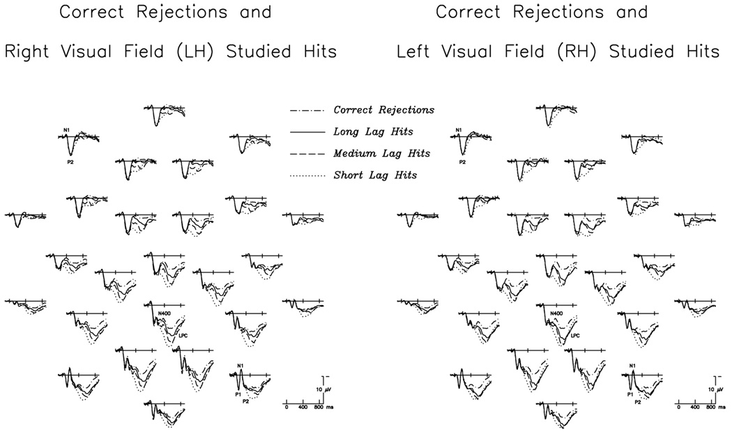
Fig. 1.
Grand average ERP waveforms to correctly identified test words, at all 26 scalp electrodes. At left are RVF/LH-studied hits (repeated at short, medium, and long lags) plotted with correct rejections; at right are LVF/RH-studied hits (repeated at short, medium, and long lags) plotted with correct rejections (note that test words appeared in central vision, and correct rejection waveforms are therefore identical in the two figures). Here, and in all subsequent figures, negative is plotted up. Latencies and amplitudes of early perceptual components (P1, N1, P2) are similar for all conditions, except for frontal P2s, which are larger for RH-studied hits. Old/new effects are visible at most electrode sites and for items studied in either VF, with greater positivity to hits than to correct rejections; this difference is strongest over centro-parietal sites and apparent in timewindows encompassing both the N400 and the LPC.

