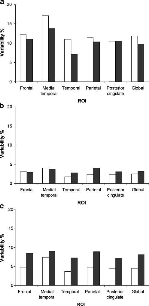
Fig. 1.
Test-retest variability is shown for a VT, b parametric SRTM2 derived BPND + 1 and c SUVr of several regions of interest (ROI): frontal, medial temporal, temporal and parietal cortex, posterior cingulate and global cortical binding. The latter is the volume-weighted average of the previously mentioned regions. In the case of VT, n = 4 and 5 for patients with Alzheimer’s disease (AD) and healthy controls, respectively; in the case of parametric SRTM2 and SUVr, n = 6 for both patient groups. AD: filled columns; healthy controls: open columns

