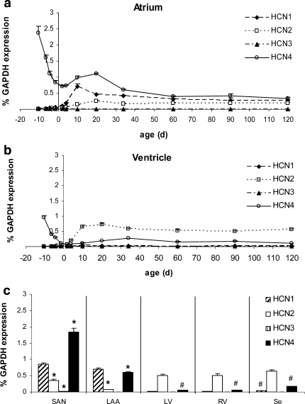
Fig. 1.
Transcription profiling of HCN isotypes throughout mouse cardiac development. a Atrial HCN isotype transcription. In the early embryonic atrium, HCN4 transcripts are the sole source of If current. HCN1-3 is absent. From birth to P34, HCN transcription is reorganized and achieves constant adult levels at P60. b Ventricular HCN isotype transcription. Only HCN4 transcripts appear in the early embryonic ventricle. Postnatally, HCN transcription is changed to predominant HCN2 and moderate HCN4 levels while HCN1 and HCN3 transcripts are virtually missing. c Regional differences at P10. In the sinoatrial node (SAN), HCN4 is expressed at a significantly higher level than in the whole atrial tissue (P < 0.05) and clearly dominates HCN1 and HCN2 transcription. In the left atrial auricle (LAA), HCN4 is reduced to one-third of the SAN level, being expressed significantly lower than in whole atrium (P < 0.05). In the left (LV) and right (RV) ventricle and in the interventricular septum (Se) HCN2 clearly dominates HCN4 while HCN1 like HCN3 is negligible. Interestingly, HCN4 is significantly higher in the Se and significantly lower in LV and RV than in the whole ventricular tissue, respectively. Statistical analysis: *P < 0.05, SAN and LAA, respectively, are compared pairwise with whole atrium at P10. #P < 0.05, LV, RV and Se, respectively, are compared pairwise with whole ventricle at P10 (n = 3)

