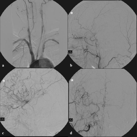
Fig. 1.
Angiogram of a 64-year-old man with bilateral ICA occlusion with collateral blood flow towards the left symptomatic hemisphere via the OphthA on the asymptomatic side, who did not suffer a recurrent ischaemic stroke during a follow-up period of 3.2 years; a bilateral ICA occlusion; b selective catheterisation of the left CCA shows filling of only a few MCA branches via the OphthA; c selective catheterisation of the right CCA shows extensive filling of ACA and MCA branches in the right hemisphere and d of the left hemisphere via the right OphthA and subsequently the AComA with filling of ACA and MCA branches

