
FIGURE 1.
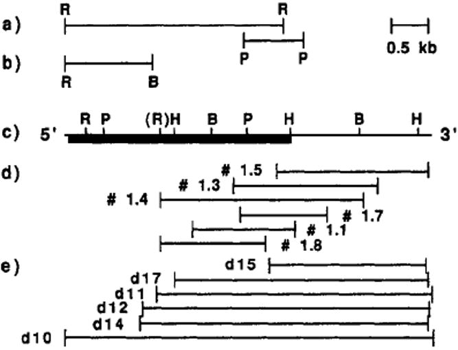
Restriction map of the probes and purified cDNAs. (a) Map of the EcoRI (R) and PstI (P) restriction fragments and EcoRI–BglII (B) fragment in (b), from human αv cDNAs used as probes for screening at low stringency. (c) Restriction map of the purified chick cDNAs. The box indicates the location of the main open reading frame. (EcoRI, R; BglII, B; PstI, P; HindIII, H). (d) Map of the six chick embryonic (E10) cDNAs (1.1, 1.3, 1.4, 1.5, 1.7, 1.8) purified with the probes mentioned in (a). (e) Map of six cDNAs (d10, d11, d12, d14, d15, and d17) purified from a chick brain (E13) cDNA library.