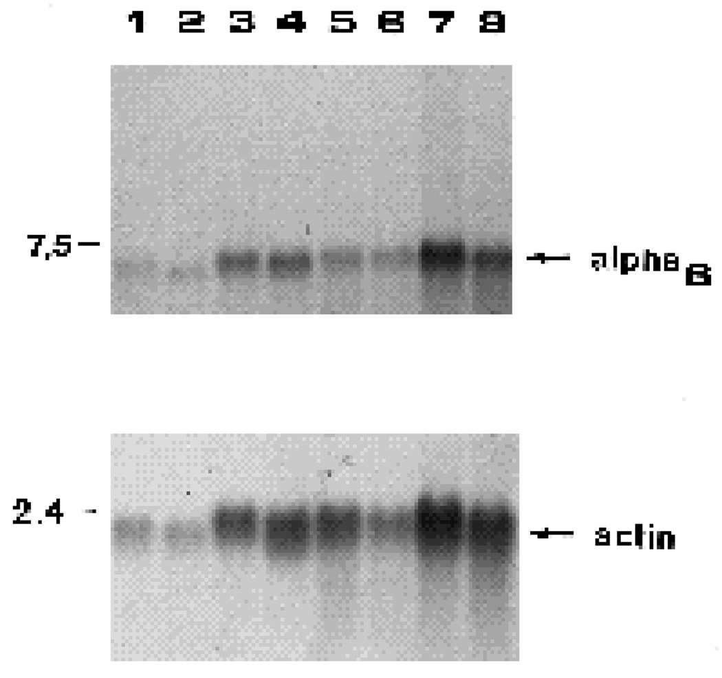
Fig. 9.
Effects of tectal ablation on integrin α6 mRNA levels in retinal ganglion cells and other retinal cells. Retinal ganglion cells (RGC) and retinal ganglion cell depleted fractions (non-RGC) were used to prepare total RNA samples. Lanes 1–4 are RNA samples from one experiment; lanes 5–8 are RNA samples from a second experiment. RGC, controls (lanes 1,5); RGC, tectum-ablated (lanes 2,6); non-RGC, controls (lanes 3,7); non-RGC, tectum-ablated (lanes 4,8). RNA samples were fractionated by agarose gel electrophoresis, transferred to nitrocellulose and incubated with 32P-α6 probe. The upper section of this blot was incubated with the 32P-α6 probe, the lower section with the 32Pactin probe. The positions of DNA standards are indicated in kb on the left. When quantitated, tectal ablation resulted in 15% and 47% increases in α6 mRNA in the RGC fractions, and 11% and 16% decreases in the non-RGC fractions respectively in the two experiments depicted here.

