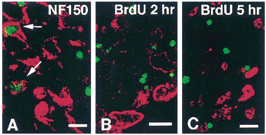
Figure 4.
Attempts to Detect Apoptosis in Neurons and Neuronal Precursors
(A) Double immunofluorescence labeling for TUNEL (green) and neurofilament (NF150, red) in a thoracic ganglion of an E11 mutant embryo. In this figure, there are two immunopositive cells for neurofilament in the field whose nuclei are labeled by the apoptosis detection method (arrows). This suggests that neurons die at this stage.
(B and C) Double labeling for TUNEL (green) and BrdU (red) after either 2 or 5 hr injection pulse of BrdU at E11 in thoracic ganglia, respectively. Colocalization was never observed, indicating that precursors are not dying in the mutant animals, but instead are lost through more rapid differentiation into cells with a neuronal phenotype. Bars, 10 µm (A and C); 10 µm (B).

