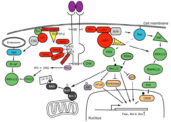
Figure 1.

Schematic diagram of Trk receptor-mediated signal transduction pathways. Binding of neurotrophins to Trk receptors leads to the recruitment of proteins that interact with specific phosphotyrosine residues in the cytoplasmic domains of Trk receptors. These interactions lead to the activation of signaling pathways, such as the Ras, phosphatidylinositol-3-kinase (PI3k), and phospholipase C (PLC)-γ pathways, and ultimately result in activation of gene expression, neuronal survival, and neurite outgrowth (see text for detailed discussions and abbreviations). The nomenclature for tyrosine residues in the cytoplasmic domains of Trk receptors are based on the human sequence for TrkA. In this diagram, adaptor proteins are red, kinase green, small G proteins blue, and transcription factors brown.
