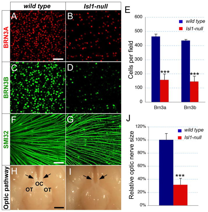
Figure 4.
Loss of RGCs in adult Isl1-null retina. (A–G) Immunostaining of whole-mount retina with anti-BRN3A (A,B), BRN3B (C,D) and SMI32 (F,G) antibodies reveals the reduction of RGCs in Isl1-null retina. (E) Quantification of BRN3A+ and BRN3B+ cells in the central retinal region of the whole-mounts (n=4 for each genotype) reveals a loss of 66% RGCs in Isl1-null retina. (H,I) Ventral views of brain show the thinner optic nerves (arrows) in Isl1-nulls. (J) Quantification of optic nerve size by measuring the cross area of H&E stained optic nerve transverse sections at the level indicated by arrows. Mean size of optic nerve in Isl1-null mice (red, n=6) is reduced to 31% of that in wild type (Blue, n=6). Each histogram represents the mean ± s.d. (***t-test P<0.001). OC, optic chiasm; OT, optic tract. Scale bar: 50 μm in A; 100 μm in F; 1 mm in H.

