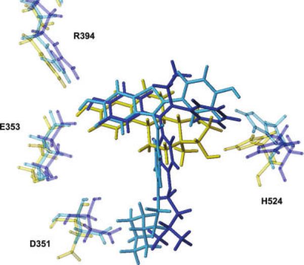
Figure 2.

Overlay of the ERα binding site complexed with 17β-estradiol (PDB code: 1ERE, yellow), 4-hydroxytamoxifen (PDB code: 3ERT, blue), and raloxifene (PDB code: 1ERR, light-blue). Only the ligands and the key interacting residues are represented in capped sticks. Glu353 and Arg394, which interact with the estradiol A ring, show a very similar behavior in the different complexes. On the contrary, His524, which interacts with the estradiol D ring, displays different orientations in different complexes, reflecting the higher degree of freedom of ligand molecules in this portion of the binding pocket. The side chain of Asp351 shows two distinct orientations, depending on the presence (SERMs) or absence (agonist) of a bulky side chain able to interact with this residue.
