Figure 2.
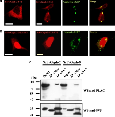
Gephyrin-specific intrabodies interact with gephyrin in mammalian cells. A. Immunofluorescence assay of the subcellular distribution of SV5-tagged anti-gephyrin intrabody (scFv-Gephyrin) ectopically expressed in HEK 293 cells in single transfection experiment (left panel) and in co-transfection with gephyrin-EGFP (right panels). ScFv-Gephyrin distribution was revealed with the anti-SV5 monoclonal antibody followed by anti-mouse TRITC-conjugated secondary antibody. Gephyrin distribution was revealed by the intrinsic green fluorescence of EGFP. B. Single (left panel) and double (right panels) transfection of the nuclear target NLS anti-Gephyrin intrabody (scFv-Gephyrin-NLS) was visualized as described in A. (Scale bar, 10 μm). C. Lysates of HEK 293 cells co-transfected with gephyrin-FLAG and scFv-Geph-2 or scFvGeph-9 were immunoprecipitated with monoclonal antibodies anti-SV5 or anti-Myc as negative control. Immunoprecipitates were analyzed by western blotting using anti-FLAG and anti-SV5 antibodies, as indicated

